

vascular
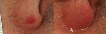 Vascular lasers and IPL for face telangiectasias have displaced other more painful and less successful alternatives. Vascular lesions that are best affordable with laser treatment if they are more visible: well-circumscribed and low flow skin lesions. The pale pink lesions that are not too different from the color of the skin are not visible to vascular lasers.
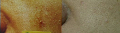 |
| Before and After two sessions Photoderm VL/PL |
Leg veins
The laser for small leg veins provides a complement but do not replace sclerotherapy. Flow of venous blood goes from thinner to thicker trough the leg veins and, from the foot to the heart. This is important in order to obtain to control the pressures of the vessels in the treatment. At the end, vessels with more pressure could be present in spite of the conventional sclerotherapy. Veins have valves that prevent blood from flowing backwards and when they are damaged or missing, it creates areas of increased pressure and consequent dilation of veins. Although the valves are not repaired new flow routes are created.
Telangiectasias, venules, varicose veins
The smaller dilated veins are called telangiectasias. They are usually red when they are more superficial and are usually not wider than 0.4 millimeters in diameter. The violet telangiectasias are superficial vessels close to 0.9 millimeters. Venules are blue-green and have a diameter of 1 to 3 millimeters. Varicose veins are thicker and bulge on the skin.
Sclerotherapy
Sclerotherapy is the most frequent non surgical technique to reduce leg veins. The sclerosant is injected into the vein and generates the closure of the vessel. It should be noted that sclerotherapy leads to a new balance between the dilated vessels and those who are not. The aim is to close the origin of the dilated vessels. Once inside, the vessel dilutes the sclerosant when it reaches larger veins and is eliminated by the body.
The course of treatment with sclerotherapy:
Results: There is no definitive cure for varicose veins and spider veins and you can get good or bad results using any treatment protocol (1). The best result is obtained when patient continues attending to the controls and doctor does not force results. Telangiectasias between 0.3 and 0.5 millimeters in diameter respond better with sclerotherapy than with the laser. Vascular lasers work better in violet vessels. Wider vessels more than 5 mm diameter with thick walls can be removed with microsurgical techniques and have more frequent complications with sclerotherapy.
Travelling: Avoid prolonged travel, 1 to 2 weeks after sclerotherapy. At high altitudes, it is not safe to proceed to sclerotherapy sessions within 1 week.
Compression stockings: It is better to use them before and after the session. Before, in order to find your best size and brand, and after because there are less chances of getting minor hematomas, less possibility of swollen ankles and less chance of spots. Symptoms of tingling in the toes force the change to a larger size. We recommended their permanent use for 72 hours after the session. During the day, take 20 to 30 mm Hg during 3 months after sclerotherapy, if possible. At night it is helpful to use an average maximum pressure of 20 mm Hg
Allergies: Allergies to sclerosant agents used in sclerotherapy (Polidocanol, Sklerol, and Ethanolamine) are very rare and the uses of products like saline and hypertonic glucose are more painful and can cause ulcers and cause spots more easily.
Bruising: They are very frequent. They resolve after 15 days. Their presence diminishes with the use of support stockings.
Matting: In some cases, we have observed the presence of smaller vessels after sclerotherapy, usually less than 0.2 mm diameter. In our experience that is caused by the use of high concentrations of the sclerosant.
Pigmentation: This complication is definitely more common at higher concentrations of sclerosant, it is more common in pigmented skins, it is seen more easily when the vessels are very wide, thick and large, thrombus are not drain properly, and compression stockings are not used for at least one month and a half which is the time that if they are not present they will not appear later. Pigmentation may occur in skin areas treated with sclerotherapy or laser. Histological examinations show hemosiderin deposits, a pigment derived from hemoglobin. Hemosiderin migrates to the dermis and to varying degrees creates an inflammatory response and leads to an increase in melanin pigmentation. Usually they resolve between 6 months and 2 years spontaneously.
Drugs: Avoid the use of minocycline and iron supplements during the treatment time.
Drainage: The most painful part of treatment. Following sclerotherapy some vessels are seen more noticeable than before, and painful to pressure. The presence of thrombi generates a perivenulitis that can persist for months and favors the extravasations of red blood cells outside of the cup if untreated.
(1) Duffy David Escleroterapia Página 71 del libro Tratamiento de las varices Editado por Murad Alam, Tri H Nguyen Elsiever SA España 2007