
skin tumors
Moles can be treated more accurately with carbon dioxide lasers and Erbium-YAG, which helps take histological examinations of the bigger ones, or evaporate the smaller ones. Some superficial lesions such as seborrheic keratosis, skin fibromas and flat warts require the use of more precise equipment to ensure a better chance of healing. Deep benign tumors can be more adequately treated with lasers to avoid larger cuts and scars.
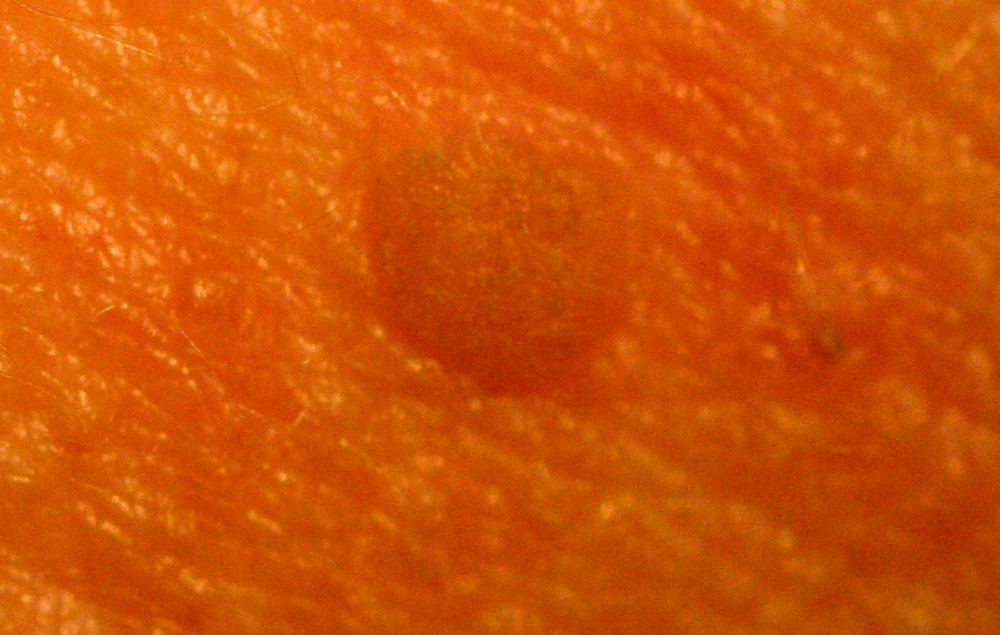
| Seborrheic Keratosis | |
 |
 |
| Before | Ablative Action of the Erbium-YAG laser |
“Acquired” melanocytic nevi
| Multiple nevi treated with CO2 and Erbium-YAG laser | |
 |
 |
| Before | After |
Most of pigmented lesions in the body come from a melanocytic origin that is, from pigment cells of the skin. Nevus grow slowly. Some are easily diagnosed, others can be confused with melanoma that is, skin cancers. Proper visualization with dermoscopy helps differentiate them from malignant lesions.
When moles are very numerous with diameters less than 2 mm, they can be vaporized with help of dermoscopy. This requires:
1. Presence of symmetry of the lesion in color, shape and structures.
2. Presence throughout the lesion, of maximum two pigmented structures and maximum two different colors (light and dark brown).
| Melanocitic nevus | |
 |
 |
| Before | CO2 laser shaving technique. Follow-up 10 years |
Lesions with diameters above 3 mm need to be treated with shaving technique with the CO2 laser for histological analysis. Most of them do not require closure with sutures, as moles are not usually wider in the deeper parts. When lesions are deep, hypertrophic scarring should be avoided shortening healing period with suture.
Moles scalp and neck skin with higher elevation are mostly superficial and can be treated with the technique of shaving with CO2 laser. Pigmented lesions require more extensive retouching but ultimately did not generate wider scars. When cutting across the thickness of the skin to make primary seals of superficial lesions, with greater possibility are raised scars. The larger incisions in the trunk thicker skin dilate with each passing month.
| Melanocytic nevus of free border of lower eyelid | |
 |
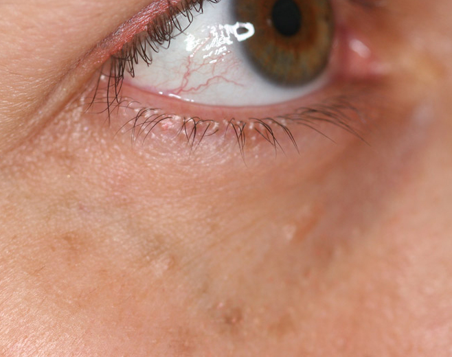 |
| Before | Result after CO2 laser |
Benign tumors of the Skin
| Fibrous papule nose | |
 |
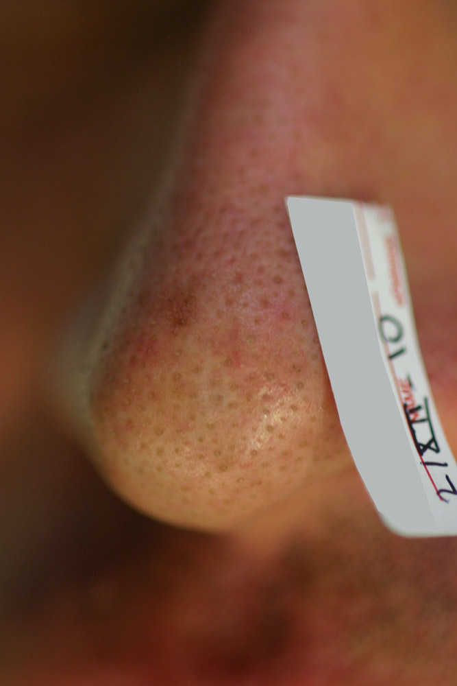 |
| Before | 10 years follow-up after laser procedure |
There are a variety of benign skin tumors that can be more properly treated with laser because incisions can be made closer to the boundaries of the lesion. If the lesion is superficial, the shaving is indicated. If lesion is deeper, the incision will be done around the closer edges making the laser cutting easier than with the thinner blade.
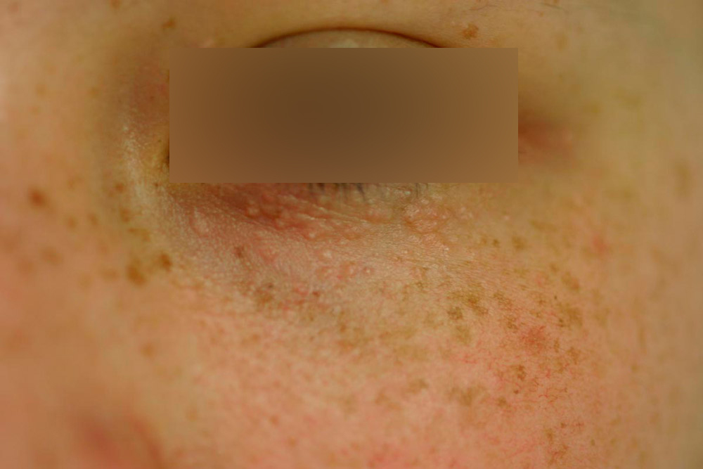
Syringomas of the eyelids
 |
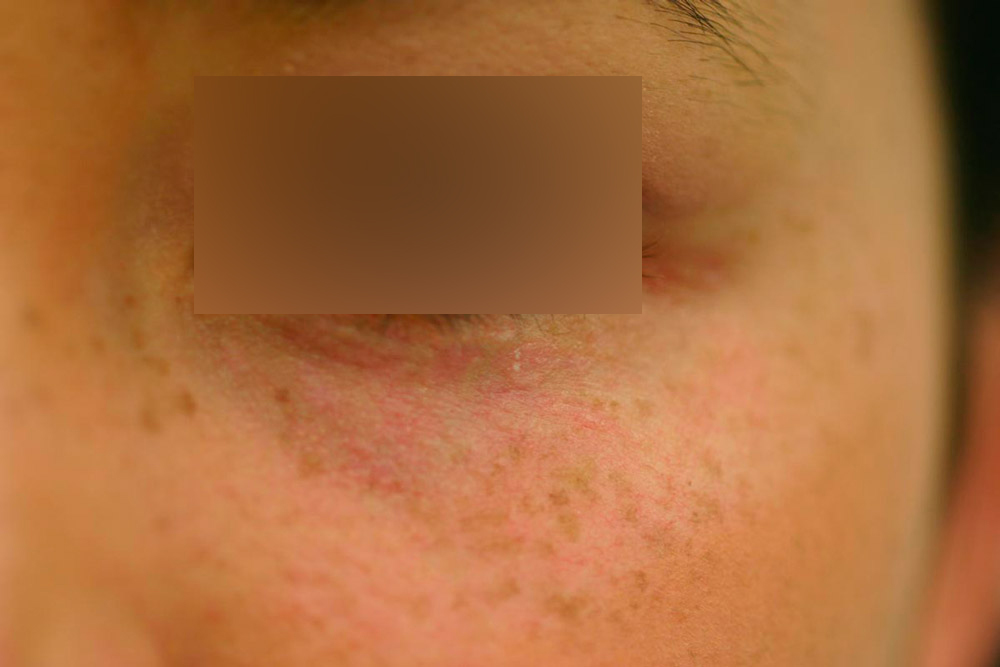 |
| Before | Laser session after 5 |
Tumor lesions such as sebaceous hyperplasia and syringomas are difficult to eradicate with instruments different than laser. The wide variety of sizes and tendency to confluence limit instruments with lower precision. Usually, deeper lesions require primary closure with simple sutures to facilitate surgery. Fractional manual technique with CO2/Erbium-YAG permits better results.
Reoperations of tumors such as epidermoid cyst is proper for the CO2 laser. The laser allows dry cuts that facilitate the dissection of the lesion including highly vascularized sites such as the scalp.
Congenital melanocytic Nevi and melanocytic spots
| Kissing / divided / Split Nevus of eyelids | |
 |
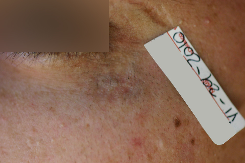 |
| Before | After 20 laser sessions |
These lesions per definition have diameters greater than 20 millimeters. They usually appear in newborn like well-circumscribed pigmented areas, which over the years may rise and develop hair. The location and extent of some congenital nevi make surgical excision difficult. The clarification of the lesions with lasers is limited to the thickness of the lesion and the persistence of the treatment by the patient. Action should be taken with different types of lasers in many sessions.
| Nevus of Ota | |
 |
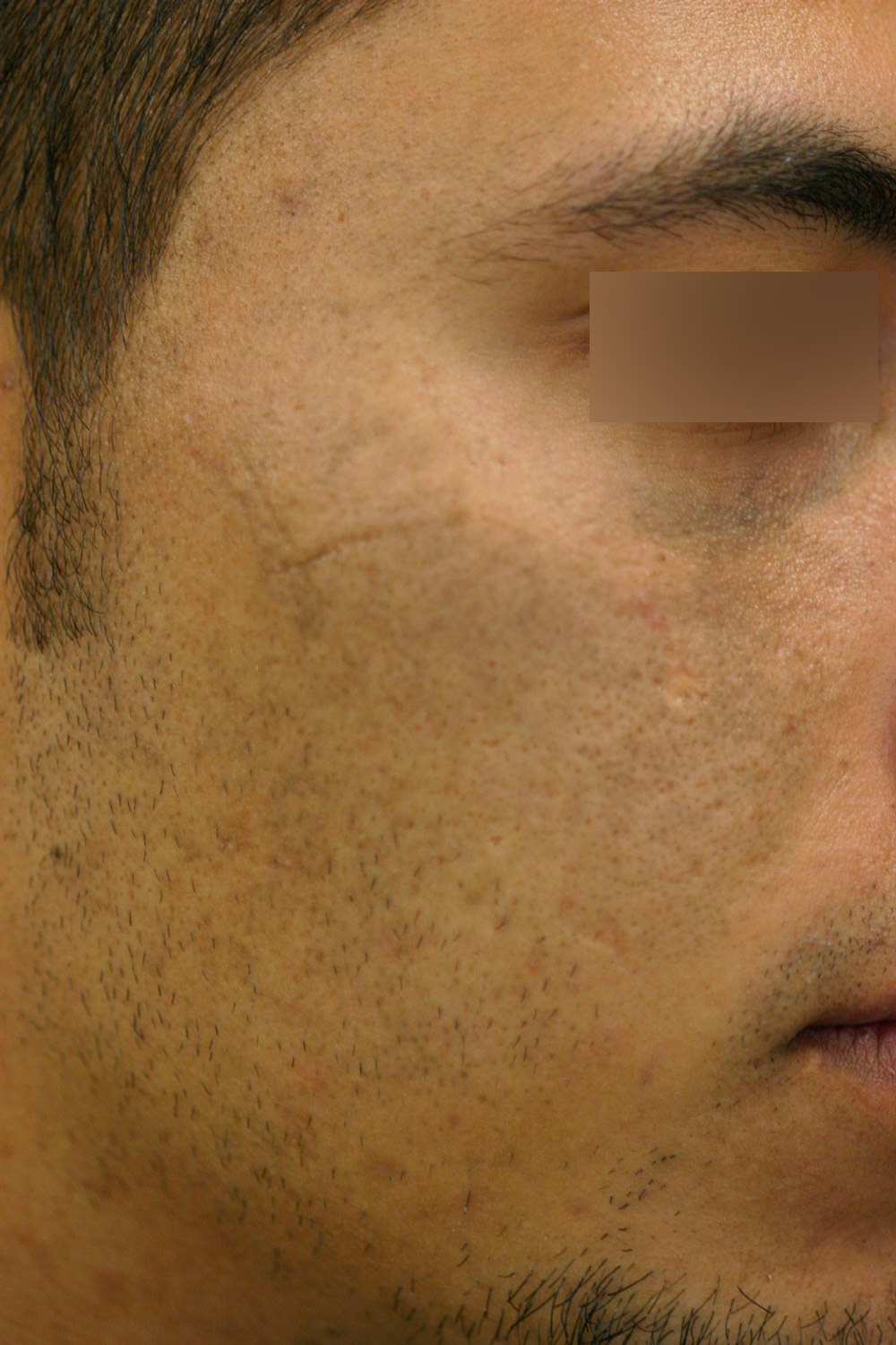 |
| Before | Result after laser treatment |
Nevus of Ota laser is presented as blue-brown spots on the forehead, eyelids and cheeks. Ota's Nevus respond to Q-Switched Nd-YAG lasers at 1064 nm because they have pigment cells loaded with melanin, so when laser fires selective damage can be done.
Nevus de Becker and Cafe au lait Spots
Nevus of Becker and Cafe au Lait spots show a higher activity of the cells to create pigment which is distributed to other epidermal cells through structures called melanosomes. Given the very wide absorption spectrum of melanin many lasers are retained in melanosomes reaching melanocytes by the heat irradiation. Other lasers have shown results based on reduction of melanocytes always them more susceptible to any aggression than other skin structures. In areas like the face Fraxel® are more comfortable because it generates no scabs. In the rest of the body we prefer Q-Switched Nd-YAG 532 with some limitations especially when dealing with very large lesions on darker skin.
| Light brown Congenital Spot | |
 |
 |
| Before | After laser treatment |